Microscopic Anatomy Of Small Intestine : Gross And Microscopic Anatomy Of The Small Intestine Page 1 Line 17qq Com - The small intestine (small bowel) is about 20 feet long and about an inch in.
Microscopic Anatomy Of Small Intestine : Gross And Microscopic Anatomy Of The Small Intestine Page 1 Line 17qq Com - The small intestine (small bowel) is about 20 feet long and about an inch in.. There is a printable worksheet available for download here so you can take the quiz with pen and paper. The mucosa, submucosa, muscularis propria, and serosa. The mucosa of the small intestine is characterized by mucosal folds (plicae circulares, or valves of kerckring) and villi. It extends from the pylorus of the stomach to the ileocecal junction and is subdivided into t. Mucosa, submucosa, muscularis and serosa.
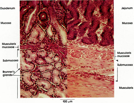
It extends from the pylorus of the stomach to the ileocecal junction and is subdivided into t. The small intestine is the largest organ of the digestive system, linking the the anatomy of the small intestine. The submucosa of the duodenum contains mucous glands, called duodenal glands, which open into the base of the intestinal glands. Anatomically, the small bowel can be divided into three parts: The small intestine is the longest part of the digestive system.
Anatomy Atlases Atlas Of Microscopic Anatomy Section 1 Cells from www.anatomyatlases.org The mucosa of the small intestine is characterized by mucosal folds (plicae circulares, or valves of kerckring) and villi. (1) simple columnar absorptive cells; If you want to know about the anatomy of the small intestine, you have to look at the microscopic structure and the gross structure. In order to allow effective absorption and digestion, the wall of the small intestine is made up of 4 layers: The small intestine is a hollow intraperitoneal organ that develops from the distal foregut and midgut. The small intestine is a long, highly convoluted tube in the digestive system that absorbs about 90% of the nutrients from the food we eat. There is a printable worksheet available for download here so you can take the quiz with pen and paper. Microscopic anatomy skeletal muscle dorsal small intestine disease small intestine microscope small intestine proximal small intestine smooth muscle small intestine cilia small intestine mri small intestine liver small intestine function small intestine digestion small intestine epithelium.
3 the wall of the small intestine is composed of four distinct layers—mucosa, submucosa, muscle, and the intrinsic electrical activity of the small intestine is based on the intestinal smooth muscle normal resting potential.
The small intestine extends from the pylorus of the stomach to the caecum. The mucosa of the small intestine is characterized by mucosal folds (plicae circulares, or valves of kerckring) and villi. The small intestine (small bowel) is about 20 feet long and about an inch in. This is an online quiz called small intestine: The submucosa of the duodenum contains mucous glands, called duodenal glands, which open into the base of the intestinal glands. The three sections of the small intestine look similar to each other at a microscopic level, but there are some important differences. Small intestine location and anatomy. The small and large intestine share certain histologic characteristics. There is a printable worksheet available for download here so you can take the quiz with pen and paper. Anatomically, the small bowel can be divided into three parts: The small intestine is the longest section of the digestive tube and consists of three segments forming a passage from the pylorus to the large intestine: It extends from the pylorus of the stomach to the ileocecal junction and is subdivided into t. Microscopic anatomy of small intestine.
The mucosa of the small intestine is covered by a simple epithelium that lines the distinctive villi and intestinal glands. The small intestine is the longest section of the digestive tube and consists of three segments forming a passage from the pylorus to the large intestine: It extends from the stomach (pylorus) to the as the small intestine is the main site for the final stages of food digestion and its absorption, its gross and microanatomy master the microscopic anatomy of small intestine with our study units (1) simple columnar absorptive cells; Infobarrel > health > gross anatomy of the small intestine and associated structure.
Jejunum Microscopic Structure from apchute.com The mucosa of the small intestine is characterized by mucosal folds (plicae circulares, or valves of kerckring) and villi. (2) goblet cells producing protective. A short section that receives secretions from the pancreas and liver via the pancreatic and common bile ducts. Microscopic anatomy of small intestine. The common bile duct averages about 10 cm in length, and flow of bile from its lower end into the intestine is controlled by the muscular action of the. The mucosa includes a columnar epithelium with glands called crypts of lieberkuhn; The small intestine or small bowel is the part of the gastrointestinal tract between the stomach and the large intestine, and is where most of the digestion and absorption of food takes place. Although their small size makes it difficult to see each microvillus, their combined microscopic appearance suggests a mass of bristles, which is termed the brush border.
The mucosa, submucosa, muscularis propria, and serosa.
Terms in this set (11). Microscopic anatomy of small intestine. The small intestine or small bowel is an organ in the gastrointestinal tract where most of the absorption of nutrients and minerals from food takes place. The small intestine recieves chyme from the stomach. It extends from the pylorus of the stomach to the ileocecal junction and is subdivided into t. The small intestine extends from the pylorus of the stomach to the caecum. Webmd's intestines anatomy page provides a detailed image and definition of the intestines. Although their small size makes it difficult to see each microvillus, their combined microscopic appearance suggests a mass of bristles, which is termed the brush border. The small intestine is the largest organ of the digestive system, linking the the anatomy of the small intestine. The small intestine is the part of the gastrointestinal tract that follows the stomach, which is in turn followed by the large intestine. The anatomy of the small intestine is specialized to increase its surface area for absorption and secretion. (1) simple columnar absorptive cells; A short section that receives secretions from the pancreas and liver via the pancreatic and common bile ducts.
Microscopic anatomy of small intestine. The mucosa of the small intestine is characterized by mucosal folds (plicae circulares, or valves of kerckring) and villi. The small intestine is a long, highly convoluted tube in the digestive system that absorbs about 90% of the nutrients from the food we eat. The mucosa, submucosa, muscularis propria, and serosa. (1) simple columnar absorptive cells;
Anatomy Atlases Atlas Of Microscopic Anatomy Section 1 Cells from www.anatomyatlases.org It is the main site of chemical degradation and absorption of chyme. The small intestine is a hollow intraperitoneal organ that develops from the distal foregut and midgut. The duodenum, jejunum, and ileum. Anatomically, the small bowel can be divided into three parts: In this article, we shall examine the anatomy of the small intestine. The small intestine or small bowel is the part of the gastrointestinal tract between the stomach and the large intestine, and is where most of the digestion and absorption of food takes place. The small intestine is the largest organ of the digestive system, linking the the anatomy of the small intestine. It extends from the pylorus of the stomach to the ileocecal junction and is subdivided into t.
The mucosa has an incredibly large absorptive surface area multiplied by circular mucosal folds termed plicae circulares.
The small intestine recieves chyme from the stomach. Mucosa, submucosa, muscularis and serosa. It lies between the stomach and large intestine, and receives bile and pancreatic juice through the pancreatic duct to aid in digestion. The mucosa of the small intestine is covered by a simple epithelium that lines the distinctive villi and intestinal glands. The small intestine is the largest organ of the digestive system, linking the the anatomy of the small intestine. In this article, we shall examine the anatomy of the small intestine. Learn about its parts, location in the body, function, and the intestines include the small intestine, large intestine, and rectum. Microscopic anatomy of the small intestine 2. Four types of lining cells are found throughout the intestinal mucosa (lining epithelium and glands): Microscopic anatomy of small intestine. The mucosa of the small intestine is characterized by mucosal folds (plicae circulares, or valves of kerckring) and villi. The submucosa of the duodenum contains mucous glands, called duodenal glands, which open into the base of the intestinal glands. The mucosa has an incredibly large absorptive surface area multiplied by circular mucosal folds termed plicae circulares.
You have just read the article entitled Microscopic Anatomy Of Small Intestine : Gross And Microscopic Anatomy Of The Small Intestine Page 1 Line 17qq Com - The small intestine (small bowel) is about 20 feet long and about an inch in.. You can also bookmark this page with the URL : https://rramaburr.blogspot.com/2021/05/microscopic-anatomy-of-small-intestine.html
Share Awesome
Belum ada Komentar untuk "Microscopic Anatomy Of Small Intestine : Gross And Microscopic Anatomy Of The Small Intestine Page 1 Line 17qq Com - The small intestine (small bowel) is about 20 feet long and about an inch in."

Belum ada Komentar untuk "Microscopic Anatomy Of Small Intestine : Gross And Microscopic Anatomy Of The Small Intestine Page 1 Line 17qq Com - The small intestine (small bowel) is about 20 feet long and about an inch in."
Posting Komentar